Case Rep Dent:海洋胶原基质载体联合可注射富血小板纤维蛋白治疗牙龈退行性缺损
2023-12-18 医路坦克 MedSci原创 发表于上海

本研究旨在检测海洋胶原基质(MCM)浸渍可注射的富血小板纤维蛋白(I- prf)和改性CAF治疗Miller 's I类和II类衰退缺陷的疗效。
牙龈萎缩缺陷在所有人群中普遍存在,并伴随一些负面的功能和美学后遗症。治疗退行性缺损的金标准治疗方法是冠状进展皮瓣(CAF)联合结缔组织移植物(CTG),其结果可预测,具有长期稳定性,且具有良好的美观性。使用CTG的主要限制是涉及到第二个手术部位,增加了患者的发病率,在治疗多发性退行性缺损时移植物组织的可用性存在问题,或者当患者的生物型较薄且手术时间增加时。
为了克服这些限制,研究了各种软组织移植替代品,可以提供与金标准相当的结果。在这里,我们使用海洋胶原基质(MCM)注入可注射的富血小板纤维蛋白(I-PRF)作为移植物替代品和CAF。

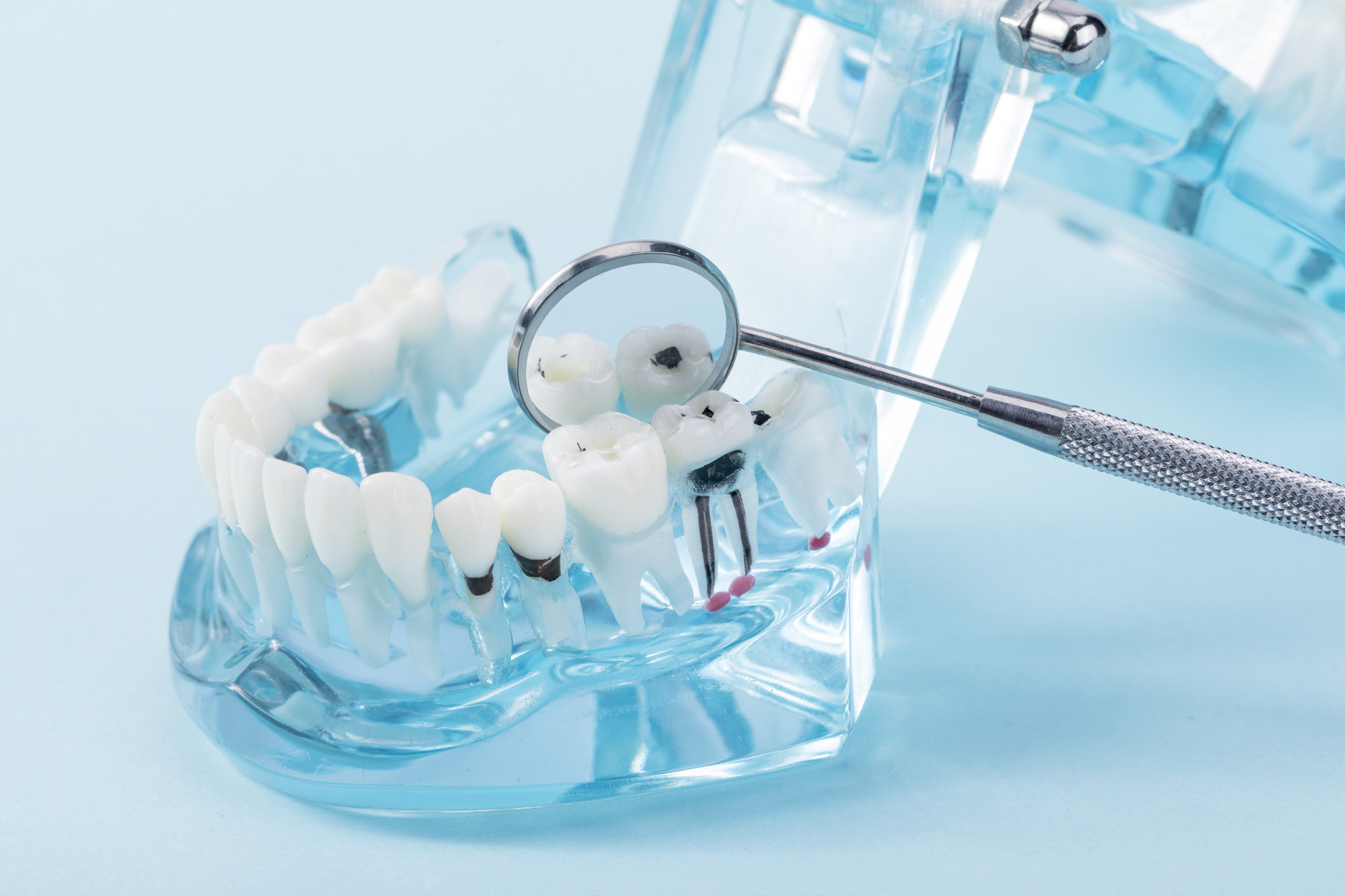
胶原蛋白是细胞外基质中的一种重要蛋白质,作为生物支架,它通过细胞的定向迁移促进血管生长,并有助于定向和有组织纤维的沉积,从而增加组织的完整性。与没有人畜共患疾病风险的陆源胶原相比,海洋胶原具有结构相似性和更好的生理生化特性。自体血小板浓缩物因其加速伤口愈合的特性已被用于医学和外科的各个领域数十年。I-PRF是Mourao等人提出的一种富含血小板的液体型纤维蛋白(II代血小板浓缩物),它比常规PRF更具优势。I-PRF可促进成纤维细胞迁移,释放生长因子水平升高,增强血管生成活性,上调创面愈合,并可促进成骨细胞迁移、粘附、增殖和分化。它对牙周病原体也有相当大的抗菌活性。牛/猪胶原基质(CM)和自体浓缩血小板作为生物替代品分别用于治疗退行性缺损取得了良好的效果,但目前还没有足够的证据证明其在退行性缺损治疗中的联合疗效。
因此,我们假设MCM具有上述优点,可以作为I-PRF在缺陷部位的载体,并且在用于牙龈萎缩缺陷的治疗时,可以共同具有促进伤口愈合和再生的协同活性。
实验描述:采用CAF + MCM + I-PRF治疗上颌10例GR缺损6例。临床参数如退退高度(RH)、退退宽度(RW)、牙根覆盖率(RC%)、附着龈宽度(WAG)、角化组织高度(KTH)、探测袋深度(PPD)、临床附着水平(CAL)、牙龈生物型(GB)、菌斑指数(PI)、视觉模拟评分(VAS-E)美学评分等进行了长达六个月的评估。
结果:在3个月和6个月的随访中观察到显著的根覆盖。
表1:研究参与者的人口统计数据。

表2:个体患者各时间点的研究参数。
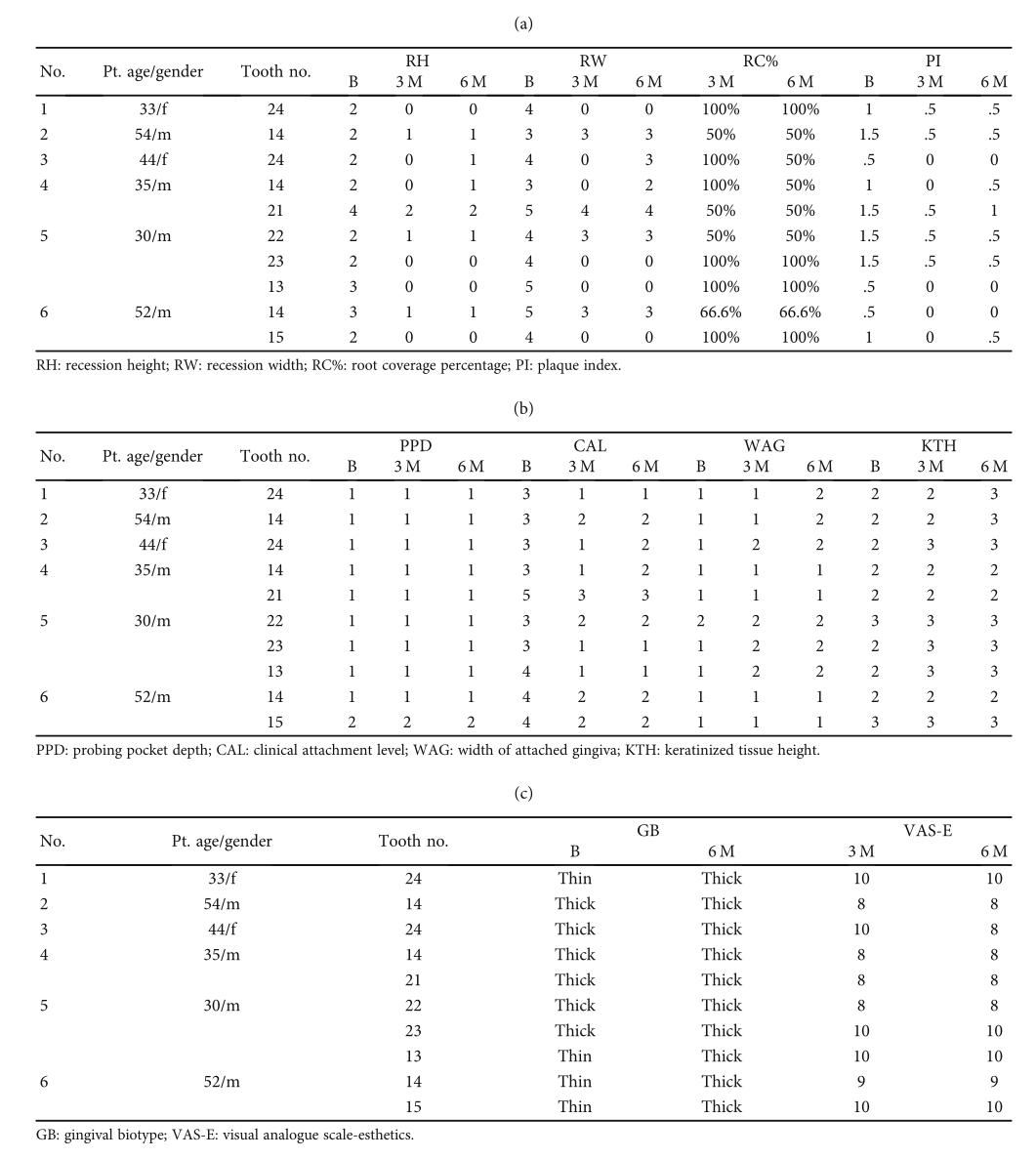
图1:I-PRF协议。(a)在指定的真空容器中从肘部静脉采集血液。(b)用Dentifuge LD C-10在60g力下700 rpm离心3分钟。(c) I-PRF分离为上液黄相。(d)用2 ml注射器提取I-PRF。
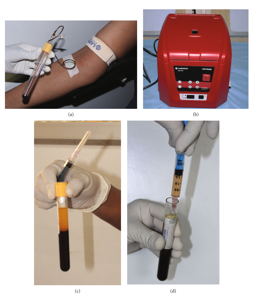
图2:基线(患者:1)。
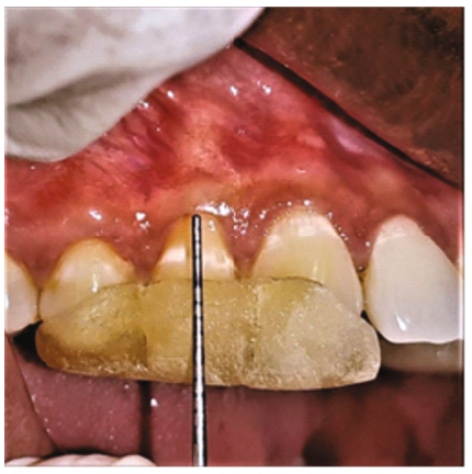
图3:皮瓣设计(患者:1)。
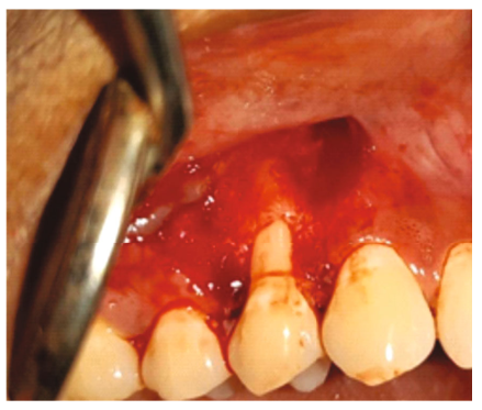
图4:I-PRF浸渍的胶原海绵在缺损部位的适应(患者:1)。
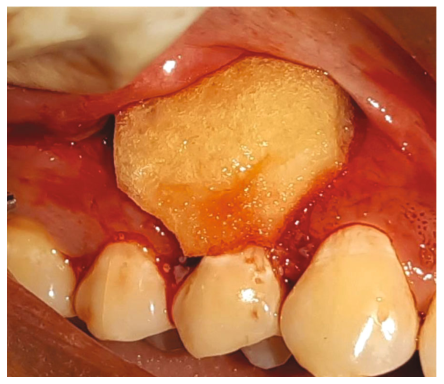
图5:皮瓣和缝合的近似(患者:1)。
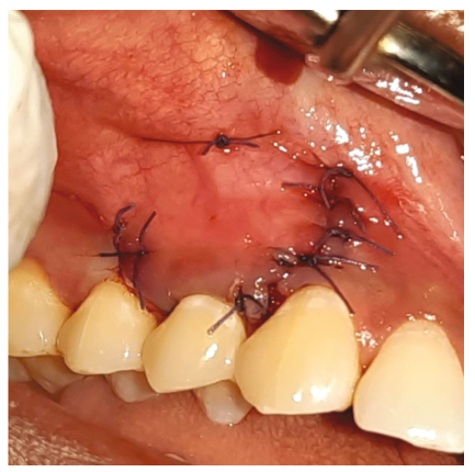
图6:基线(患者:6)。
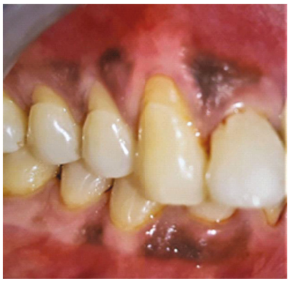
图7:皮瓣设计(患者:6)。

图8:I-PRF浸渍的胶原海绵在缺损部位的适应(患者:6)。
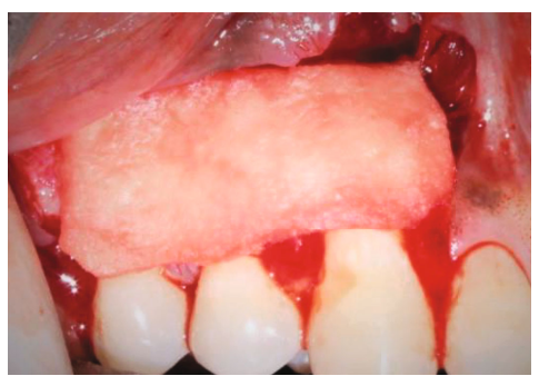
图9:皮瓣和缝合的近似(患者:6)。
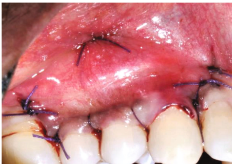
图10:6个月随访(患者:1)。
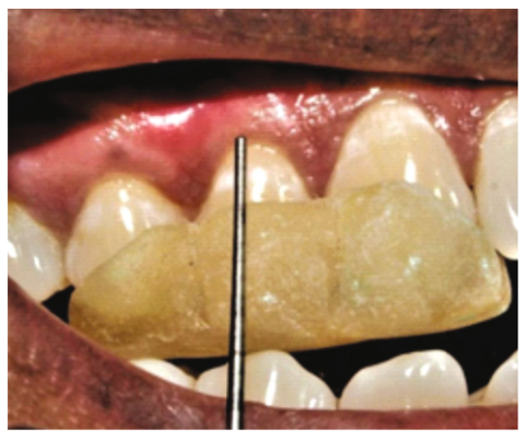
图11:随访6个月(患者:6)。

图12:基线与术后6个月照片(患者:6)。

表3:各时间点的研究参数

表4:参数在时间间隔内的变化

结论:所提出的治疗方法在管理GR缺陷和改善软组织生物型方面是有效的,而没有与软组织收获相关的发病率。
原始出处:
 Case Rep Dent:海洋胶原基质载体联合可注射富血小板纤维蛋白治疗牙龈退行性缺损
 分享
分享
本网站所有内容来源注明为“梅斯医学”或“MedSci原创”的文字、图片和音视频资料,版权均属于梅斯医学所有。非经授权,任何媒体、网站或个人不得转载,授权转载时须注明来源为“梅斯医学”。其它来源的文章系转载文章,或“梅斯号”自媒体发布的文章,仅系出于传递更多信息之目的,本站仅负责审核内容合规,其内容不代表本站立场,本站不负责内容的准确性和版权。如果存在侵权、或不希望被转载的媒体或个人可与我们联系,我们将立即进行删除处理。
在此留言


#富血小板纤维蛋白# #海洋胶原基质# #牙龈退行性缺损#
117 举报